Hair loss is a problem that affects millions of people worldwide. Although there are some treatments currently available, none represent a definitive cure. However, there are some drugs that are in advanced clinical trials and could represent a solution to the problem of hair loss.
Hair loss affects millions around the world, but a ray of hope shines through recent developments. Meet Clascoterone, a potential game-changer in the fight against Androgenetic Alopecia (AGA). Although still in clinical trials, Clascoterone shows immense potential in the treatment of hair loss, possibly surpassing existing treatments like minoxidil.
Unlocking the future:
Clascoterone
[CB-03-01] is a topical medication initially designed for the treatment of
acne. This molecule is a potent androgen receptor inhibitor, so its potential
to combat androgenetic alopecia is being tested.
Androgenetic
alopecia is characterized by an increased effect of testosterone on androgen
receptors in dermal papilla cells. By blocking these receptors, testosterone
will not have a negative effect on follicle cells, preventing miniaturization
and hair loss.
Outlining the path ahead:
As phase III trials begin, Clascoterone has significant potential to revolutionize the treatment of hair loss. These trials aim to evaluate its efficiency and safety in the treatment of AGA, a common but challenging condition that affects millions worldwide. If approved by the FDA, Clascoterone would be the first effective topical drug found against androgenetic alopecia in nearly three decades!
While the path to fight hair loss is ongoing, the progress of Clascoterone is a beacon of hope. At CapilarFix®, we remain vigilant, combining science and experience to transform lives affected by hair loss.
Schedule your consultation today!
References:
Dhillon S. Clascoterone: First Approval. Drugs. 2020 Nov;80(16):1745-1750. doi: 10.1007/s40265-020-01417-6. PMID: 33030710. H. Y. Sun, D. F. Sebaratnam, Clascoterone as a novel treatment for androgenetic alopecia, Clinical and Experimental Dermatology, Volume 45, Issue 7, 1 October 2020, Pages 913–914, https://doi.org/10.1111/ced.14292
https://capilarfix.com/wp-content/uploads/2020/04/logo-web-lowres-287x120-1.png00PhD. Raquel Cuevas-Díaz Duránhttps://capilarfix.com/wp-content/uploads/2020/04/logo-web-lowres-287x120-1.pngPhD. Raquel Cuevas-Díaz Durán2024-04-17 03:04:122024-04-17 03:04:13Know Clascoterone and how it could fight Androgenetic Alopecia
Hair loss is a problem that affects millions of people worldwide. Although there are some treatments currently available, none represent a definitive cure. However, a recent study conducted by researchers at Northwestern University may change this in the future.
The study, published in the journal PNAS, found that age-related stiffness in hair follicle stem cells can inhibit hair growth. By increasing the production of a micro-RNA called miR-205, researchers were able to make these stem cells more flexible, promoting hair growth in both young and old mice.
Implications of the study
The findings of this study have important implications for the search for new treatments for baldness. Some key points:
It indicates that it is possible to stimulate hair growth by manipulating the mechanical properties of hair follicle stem cells.
It opens the possibility of developing new treatments that promote hair growth by making these stem cells more flexible.
Researchers plan to test whether topically administered miR-205 can stimulate hair growth in humans.
If these assays are successful, they could facilitate the development of new effective therapies for baldness.
What will be the future of baldness treatments?
Although several years of research are still needed before a stem cell-based and micro-RNA treatment for baldness is available on the market, this study represents a significant scientific breakthrough.
It demonstrates that manipulating hair follicle stem cells is a promising strategy and opens the door to the development of new therapies that can effectively and durably promote hair growth.
In conclusion, baldness remains a troublesome problem for millions, but the future looks promising. If future studies in humans confirm the initial findings, we may have new stem cell-based treatments for hair loss in a few years.
At CapilarFix®, we are aware of these scientific advances because our mission is to stop each patients’ hair loss by combining science and experience. Let’s continue to hope for a future with more hair!
Alopecia areata is a condition in which the immune system attacks an individual’s own hair follicles, causing hair loss. The key players in this story are regulatory T cells, whose role has been studied for a long time in the context of autoimmune diseases.
From immunosuppression to regeneration:
However, a study conducted by scientists from
the Salk Institute revealed that these immune
cells, called Regulatory T cells, are not only immunosuppressive but also
interact with skin cells using a hormone to promote hair growth and
regeneration.
These unexpected hormones are glucocorticoid
hormones, derived from cholesterol and produced in various parts of the body.
They are used by regulatory T cells as
messengers to stimulate hair regeneration.
This process is a revelation in the field of hair medicine, as it was
believed that these cells only suppressed the immune response.
Research at the Salk Institute
Scientists at the Salk Institute began their research without the intention of
studying hair loss. However, when they studied mice with glucocorticoid
receptors and induced hair loss, they observed surprising results.
Mice with glucocorticoid receptors experienced significant hair growth
compared to those lacking these receptors.
This discovery opened up new perspectives in
the treatment of alopecia and other conditions related to hair loss.
In cases of acute alopecia, we now know that
applying glucocorticoids also has the additional benefit of stimulating the production of TGF-3 by the skin’s
regulatory T cells,
which promotes the activation of hair follicle stem cells.
Now you know, at CapilarFix, we take pride in staying at the forefront of hair research. We are committed to improving the quality of life of our patients and harnessing these scientific advances for the benefit of those struggling with hair loss.
References:
[1] Glucocorticoids initiate regulatory T cell and stem-cell crosstalk to grow new hair. Nat Immunol 23, 1006–1007 (2022). https://doi.org/10.1038/s41590-022-01250-x
https://capilarfix.com/wp-content/uploads/2020/04/logo-web-lowres-287x120-1.png00PhD. Raquel Cuevas-Díaz Duránhttps://capilarfix.com/wp-content/uploads/2020/04/logo-web-lowres-287x120-1.pngPhD. Raquel Cuevas-Díaz Durán2024-02-07 10:10:002024-03-07 21:43:14The immune system and the hair follicle
Stem Cells in Hair Follicles: Bulb and Outer Bulb, The Hair Clock and Its Impact on Hair Growth, Stem Cell Dysregulation and Hair Disorders, Future Advances: Stem Cell-Based Hair Therapies.
The science behind hair growth and related conditions has evolved tremendously in recent years. In this article, we will explore a fundamental component: stem cells in hair follicles. These cells, present in two subpopulations, play a crucial role in the hair cycle and their dysregulation underlie the onset of hair diseases.
Stem Cells in Hair Follicles: Bulb and Outer Bulb
Within hair follicles, two types of stem cells can be found: outer bulb stem cells and dermal papilla stem cells. The former reside in the outer bulb, while the latter are located in the inner bulb. These stem cells play a leading role in generating new hair, as they coordinate the hair cycle and give rise to the cells that form the hair.
Microbiota varies from person to person and even within different body parts. We know that the hair follicle, especially the infundibulum is highly rich in microbiota and immune system cells. Alterations in microbiota concentrations and distributions may cause inflammation and damage to the infundibulum, deriving potentially in hair pathologies.
The Hair Clock and Its Impact on Hair Growth
The hair clock is a metaphor used to describe the hair cycle and consists of the anagen, catagen, and telogen phases. During the anagen phase, stem cells in the bulb become active, leading to hair growth. As the cycle progresses, these cells enter a resting period in the catagen phase, and finally, in the telogen phase, mature hair is shed, restarting the cycle.
Stem Cell Dysregulation and Hair Disorders
Precise communication between stem cells and other cell populations is essential. Dysregulation of this communication can lead to hair disorders and trigger hair loss. Understanding these imbalances is crucial for developing effective treatments.
Future Advances: Stem Cell-Based Hair Therapies
The constantly expanding knowledge of stem cells in hair follicles has opened exciting doors in hair therapy. Scientists are exploring how to manipulate these stem cells to boost hair growth in individuals with hair issues. Stem cell therapy and tissue engineering offer promising solutions to address alopecia and baldness.
Stem cells in hair follicles are crucial pieces in the puzzle of hair growth and related diseases. The hair clock guides the hair cycle, while dysregulation can trigger hair problems. As we advance in understanding these processes, stem cell-based therapies present a promising future in the fight against alopecia and baldness.
At CapilarFixTM we are experts at taking care of your hair follicles and your hair. We can recommend the best products that will help you maintain a balanced capilar microbiota.
https://capilarfix.com/wp-content/uploads/2020/04/logo-web-lowres-287x120-1.png00PhD. Raquel Cuevas-Díaz Duránhttps://capilarfix.com/wp-content/uploads/2020/04/logo-web-lowres-287x120-1.pngPhD. Raquel Cuevas-Díaz Durán2024-01-31 10:10:002024-03-07 21:42:59Stem Cells in Hair Follicles: The Hair Clock and Its Influence on Hair Health
The human body is colonized by trillions of microorganisms, collectively referred to as “microbiota”. In humans, microbiota inhabits diverse locations in our body such as the mouth, nasal cavities, skin, gastrointestinal tract, urogenital tract, respiratory tract, vagina, and other places. Due to advances in sequencing technologies in the last decade, a huge amount of research has demonstrated an important role of human microbiota in health and disease, particularly gut microbiota [1]. Here we will focus in hair follicle microbiota.
Human skin and hair follicles host a vast diversity of microorganisms including bacteria, viruses, fungi, and mites [2]. Microorganisms can be beneficial or pathogenic and the establishment of an equilibrium is crucial for health and disease of our skin and hair. Fortunately, the great majority of our skin and hair follicle residents are non-pathogenic and probably contribute to homeostasis by maintaining a constant dialogue with our immune system. However, stress and a variety of environmental factors are associated with microbiota imbalance.
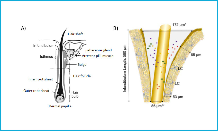
Approximately 100,000 – 150,000 hair follicles are located in the scalp. Hair follicles are colonized by unique and complex microbiota. Fungi strain Malassezia and bacterial strains Propiobacterium, Cutibacterium, and Staphylococcus are among the most abundant microorganisms in human scalps. Compared to the skin, the hair follicle favors microbial growth because it is moist, well-perfused, relatively UV-protected and has a less acidic pH [3]. The hair follicle is an organ found in skin. There are three major segments of hair follicles: lower, middle and upper segments (Figure 1). The infundibulum covers most of the upper segment. The infundibulum is the funnel-shaped uppermost portion of the follicle. It is typically filled with sebum and debris. Due to its location, the infundibulum is a major interface zone between our skin and the environment, making it a microbiota rich place. Not surprisingly, the infundibulum is endowed with a specialized immune system. However, diverse studies have demonstrated that the infundibulum is involved in skin diseases such as acne, infundibular folliculitis, cysts, hidradenitis suppurative, keratosis pilaris, and a subtype of basal cell carcinoma [4]. An imbalance in the microbiota impacts the immune system and both are key elements in the pathogenesis of chronic scalp diseases.
Figure 1: A) Parts of a hair follicle. The infundibulum is the uppermost part of the hair follicle and is in contact with the environment. B) Zoom-in of the infundibulum, a funnel-shaped region rich in microbiota. Diverse microorganisms are depicted in colors. Figure adapted from [10].
A typical example of how alterations in the composition of hair follicle microbiota may impact diseases is observed in dandruff, a mild type of seborrheic dermatitis. Studies have shown that dandruff and seborrheic dermatitis are correlated with alterations in bacterial and fungal microbiota [5-8]. Similarly, microinflammation and infiltration of mononuclear cells and lymphocytes have been observed in infundibulum samples of patients with androgenetic alopecia [9]. However, further studies are needed to understand the relationship between the interactions of microbiota, hair follicle cells, and host immune system.
Microbiota varies from person to person and even within different body parts. We know that the hair follicle, especially the infundibulum is highly rich in microbiota and immune system cells. Alterations in microbiota concentrations and distributions may cause inflammation and damage to the infundibulum, deriving potentially in hair pathologies.
At CapilarFixTM we are experts at taking care of your hair follicles and your hair. We can recommend the best products that will help you maintain a balanced capilar microbiota.
References:
[1] Gilbert JA,
Blaser MJ, Caporaso JG, Jansson JK, Lynch SV, Knight R, Current understanding
of the human microbiome. Nat Med. 2018; 24: 392-400
[2] Oh, J.,
Byrd, A. L., Deming, C., Conlan, S., NISC Comparative Sequencing Program, Kong,
H. H., & Segre, J. A. (2014). Biogeography and individuality shape function
in the human skin metagenome. Nature, 514(7520), 59–64.
[3] Lousada, M., Lachnit, T.,
Edelkamp, J., Rouillé, T., Ajdic, D., Uchida, Y., Di Nardo, A., Bosch, T. and
Paus, R. (2020), Exploring the human hair follicle microbiome. Br J Dermatol. Accepted Author
Manuscript. doi:10.1111/bjd.19461
[4] Schneider, M. R., &
Paus, R. (2014). Deciphering the functions of the hair follicle infundibulum in
skin physiology and disease. Cell and tissue research, 358(3),
697–704.
[5] C. Clavaud, R. Jourdain, A. Bar‐Hen, M. Tichit, C. Bouchier, F. Pouradier, C. El Rawadi, J. Guillot, F. Menard‐Szczebara, L. Breton, J. P. Latge, I. Mouyna, Dandruff is associated with disequilibrium in the proportion of the major bacterial and fungal populations colonizing the scalp, PLoS ONE 2013, 8, e58203.
[6] L. Wang, C. Clavaud, A. Bar‐Hen, M. Cui, J. Gao, Y. Liu, C. Liu, N. Shibagaki, A. Gueniche, R. Jourdain, K. Lan, C. Zhang, R. Altmeyer, L. Breton, Characterization of the major bacterial-fungal populations colonizing dandruff scalps in Shanghai, China, shows microbial disequilibrium, Exp. Dermatol. 2015, 24, 398.
[7] R. C. Soares, P. H. Camargo‐Penna, V. C. de Moraes, R. De Vecchi, C. Clavaud, L. Breton, A. S. Braz, L. C. Paulino, Dysbiotic Bacterial and Fungal Communities Not Restricted to Clinically Affected Skin Sites in Dandruff, Front Cell. Infect. Microbiol. 2016, 6, 157.
[8] T. Park, H. J. Kim, N. R. Myeong, H. G. Lee, I. Kwack, J. Lee, B. J. Kim, W. J. Sul, S. An, Collapse of human scalp microbiome network in dandruff and seborrheic dermatitis, Exp. Dermatol. 2017, 26, 835.
[9] Mahé, Y. F., Michelet, J.
F., Billoni, N., Jarrousse, F., Buan, B., Commo, S., Saint-Léger, D., &
Bernard, B. A. (2000). Androgenetic alopecia and microinflammation. International
journal of dermatology, 39(8), 576–584.
[10] Pisal, Rishikaysh & Dev, Kapil & Diaz, Daniel
& Shaikh Qureshi, Wasay Mohiuddin & Filip, Stanislav & Mokrý,
Jaroslav. (2013). Signaling Involved in Hair Follicle Morphogenesis and
Development. International journal of
molecular sciences. 15. 1647-70.
Vitamin D plays an important role in calcium homeostasis and bone health. It has been demonstrated that vitamin D is also associated with several autoimmune diseases such as lupus, type I diabetes mellitus, rheumatoid arthritis, multiple sclerosis, vitiligo, and psoriasis among others [Hewison, 2012]. Deficiencies in vitamin D may derive in abnormal autoimmunity responses [Arnson, 2007]. Furthermore, vitamin D and its receptor have been implicated in the pathogenesis of diverse forms of hair loss [Conic et al., 2018].
We obtain a small amount of vitamin D from our daily diet, however most of the vitamin D in our body is synthesized by keratinocytes in the epidermis after exposure to solar radiation. Vitamin D acts in human body cells through the vitamin D receptor (VDR), triggering a signaling cascade which modulates the transcription of target genes involved in regulating homeostasis. VDR is a nuclear receptor expressed in numerous cells and tissues in the human body including the skin. VDR is expressed in the two major cell populations conforming the hair follicle: epidermal keratinocytes and dermal papilla cells. Studies have demonstrated that absence of VDR leads to hair loss by hair cycle dysregulation [Mady et al., 2016].
The first evidence suggesting a role of VDR in hair cycle was derived from the observation of alopecia in patients with type IIA vitamin D-dependent rickets [Brooks et al., 1978]. Vitamin D-dependent rickets type IIA is a disorder in bone formation caused by a defect in the VDR gene. Interestingly most of the patients with this disorder also had alopecia. However, the molecular mechanisms by which vitamin D and its receptor VDR regulate hair cycle are still unknown.
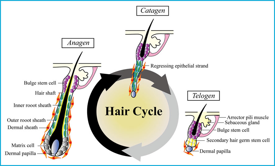
Hair regeneration depends on the activation of hair follicle stem cells. The hair follicle is an organ that undergoes cyclic involution and regeneration throughout life [Paus et al., 1999]. The hair cycle consists in three cycling phases: anagen (growth), catagen (involution), and telogen (resting). In the telogen phase, the dermal papilla and the bulge come closer together and this approximation allows signaling between the dermal papilla and keratinocyte stem cells in the bulge, inducing a new proliferative anagen phase [Paus et al., 1999] as observed in the next Figure.
Figure: Hair cycle phases anagen (growth), catagen (involution) and telogen (resting). [Chen et al., 2020]
The expression of VDR is increased in the hair follicle during the late anagen and catagen phases of the hair cycle; correlating with decreased proliferation and increased differentiation of keratinocyte stem cells [Demay et al., 2007]. VDR is essential for the start of a new anagen phase. Several mutations have been found which may interfere with VDR’s function increasing the risk of hair loss [Simon et al., 2010].
Additionally, levels of serum vitamin D have also been correlated to hair loss. Diverse studies have demonstrated insufficiency or significantly lower serum levels of vitamin D in patients with Alopecia Areata as compared to healthy controls [Yilmaz et al., 2012; Aksu et al., 2014; Ghafoor et al., 2017; Gade et al., 2018]. Supplementation with vitamin D has also been observed to protect hair follicles from chemotherapy-induced alopecia [Wang et al., 2006; Jimenez et al., 1996].
References:
Hewison M. An update on vitamin D and human immunity. Clin Endocrinol (Oxf). 2012;76:315–25.
Arnson Y, Amital H, Shoenfeld Y. Vitamin D and autoimmunity:
new aetiological and therapeutic considerations. Ann Rheum Dis. 2007;66:1137–42.
Conic, R., Piliang, M., Bergfeld, W., &
Atanaskova-Mesinkovska, N. (2018). Vitamin D Status in Scarring and
Non-Scarring Alopecia. Journal of the American Academy of Dermatology,
S0190-9622(18)30631-5.
Mady, L. J., Ajibade, D. V., Hsaio, C.,
Teichert, A., Fong, C., Wang, Y., Christakos, S., & Bikle, D. D. (2016).
The Transient Role for Calcium and Vitamin D during the Developmental
Hair Follicle Cycle. The Journal of investigative dermatology, 136(7),
1337–1345.
Brooks, M. H., Bell, N. H., Love, L., Stern, P.
H., Orfei, E., Queener, S. F., Hamstra, A. J., & DeLuca, H. F. (1978).
Vitamin-D-dependent rickets type II. Resistance of target organs to
1,25-dihydroxyvitamin D. The New England journal of medicine, 298(18),
996–999.
Paus, R., & Cotsarelis, G. (1999). The
biology of hair follicles. The New England journal of medicine, 341(7),
491–497.
Demay, M. B., MacDonald, P. N., Skorija, K.,
Dowd, D. R., Cianferotti, L., & Cox, M. (2007). Role of the vitamin D
receptor in hair follicle biology. The Journal of steroid biochemistry and
molecular biology, 103(3-5), 344–346.
Simon, K. C., Munger, K. L., Xing Yang, &
Ascherio, A. (2010). Polymorphisms in vitamin D metabolism related genes and
risk of multiple sclerosis. Multiple sclerosis
(Houndmills, Basingstoke, England), 16(2),
133–138.
Yilmaz N, Serarslan G, Gokce C. Vitamin D
concentrations are decreased in patients with alopecia areata. Vitam Miner. 2012;1:105–109.
Aksu Cerman A, Sarikaya Solak S, Kivanc Altunay
I. Vitamin D deficiency in alopecia areata. Br J Dermatol. 2014;170:1299–1304.
Ghafoor R, Anwar MI. Vitamin D deficiency in
alopecia areata. J Coll Physicians Surg
Pak. 2017;27:200–202.
Gade VKV, Mony A, Munisamy M, Chandrashekar L,
Rajappa M. An investigation of vitamin D status in alopecia areata. Clin Exp Med. 2018;18:577–584.
Wang J, Lu Z, Au JL. Protection against
chemotherapy-induced alopecia. Pharm Res.
2006;23:2505–14.
Chen, C., Huang, W., Wang, E.H.C. et al. Functional complexity of hair follicle stem cell niche and therapeutic targeting of niche dysfunction for hair regeneration. J Biomed Sci 27,43 (2020).
https://capilarfix.com/wp-content/uploads/2020/04/logo-web-lowres-287x120-1.png00PhD. Raquel Cuevas-Díaz Duránhttps://capilarfix.com/wp-content/uploads/2020/04/logo-web-lowres-287x120-1.pngPhD. Raquel Cuevas-Díaz Durán2020-09-17 18:13:252024-03-07 21:42:30Vitamin D deficiency and hair loss
We are currently in the midst of a pandemic situation
orchestrated by the SARS-CoV2 (COVID-19) virus. The name SARS stands for Severe
Acute Respiratory Syndrome and CoV2 indicates that the virus belongs to the
family of Coronaviridae.
Coronaviruses are characterized for having a surface covered with protrusions
called spikes, giving them a crown-like appearance. Genomic sequence analysis
suggest that the virus originated through natural selection in bats or Malayan
pangolins and then jumped into humans, a process called “zoonotic transfer” [Lam
et al., 2020].
Early epidemiologic reports have shown a disproportionate prevalence of COVID-19 severe cases between adult women and men (42% in males vs 58% in females) [Shi et al., 2020]. Furthermore, a bigger difference is observed in the number of severe COVID-19 cases between pre-pubescent children and adults [Lu et al., 2020]. In children, only 0.6% of COVID-19 cases are severe. In Mexico epidemiological reports are updated daily by Secretaria de Salud. Interestingly, as of July 22, 2020 only 34.8% of COVID-19 deaths were of women whereas 65.18% were men [https://coronavirus.gob.mx/]. An explanation for the skewed prevalence of severe COVID-19 cases between males and females is still unknown.
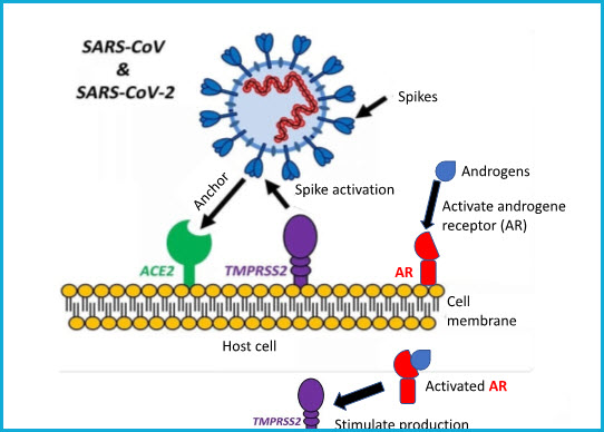
SARS-CoV-2 virus uses its spikes to target and anchor cells to infect. The main virus’ entry point in a host cell is a protein called ACE2. ACE2 is found on the surface of cells from a variety of human organs such as lungs, arteries, heart, kidneys, and intestines. Spikes need to be activated, process known as “priming”, for anchoring host cells. Spike activation is performed by the TMPRSS2 protein, also found on the surface of host cells [Hoffman et al., 2020]. Interestingly, when the androgenetic receptor (AR) is activated, it stimulates the production of TMPRSS2 protein which in turn activates more SARS-CoV2 spikes. Similarly, as in hair follicles, the androgenetic receptor (AR) requires of hormones called androgens (e.g. testosterone) to become activated. The next figure depicts the SARS-CoV2 molecular infection mechanism when encountered with a host cell.
Figure 1 – SARS-CoV2 infection mechanisms. Adapted from Hoffman en al., 2020.
In a study performed between March 23 and April 12, 2020 in three hospitals of Madrid, Spain, dermatologists classified 175 COVID-19 patients as “no alopecia”, “moderate androgenetic alopecia”, or “severe androgenetic alopecia” prior to hospitalization. Results demonstrate that 67% of the patients depict symptoms of moderate or severe androgenetic alopecia. Additionally, 79% of COVID-19 severe cases had androgenetic alopecia as well [Wambier et al., 2020]. These numbers show a clear trend; however, further epidemiological studies are required to assess a correlation between the degree of androgenetic alopecia and the severity of COVID-19 infection. It is worth mentioning that clinical trials highlighting the use of anti-androgen drugs are currently ongoing. These drugs aim at reducing androgen levels, thus decreasing the activation of androgenetic receptors in COVID-19 patients.
All these
observations have led scientists to study the potential correlation between
androgenetic alopecia with the severity of COVID-19 infection under the
hypothesis that places androgens as the main cause of COVID-19 severity. The
potential correlation between androgenetic alopecia and COVID-19 has been termed
the “Gabrin Sign” in honor of Doctor Frank Gabrin, the first American physician
who died of COVID-19 [Wambier et al., 2020]. Interestingly, Doctor Gabrin
suffered from androgenetic alopecia and was victim of a severe COVID-19
infection.
At CapilarFix® we are
not experts in COVID-19 however we are skilled in treating alopecia. Perhaps
(we can’t be sure yet), by treating androgenetic alopecia we could indirectly
be lowering the risk of severe COVID-19 infection.
References:
Lam, T.T., Jia, N., Zhang, Y. et al. Identifying SARS-CoV-2-related coronaviruses in Malayan pangolins. Nature 583,282–285 (2020). https://doi.org/10.1038/s41586-020-2169-0
Shi Y., Yu X., Zhao H., Wang H., Zhao R., Sheng
J. Host susceptibility to severe COVID-19 and establishment of a host risk
score: findings of 487 cases outside Wuhan. Crit Care. 2020;24(1):108.
Lu X, Zhang L, Du H, et al. SARS‐CoV‐2
infection in children. N Engl J Med. 2020.
Hoffmann M., Kleine-Weber H., Schroeder S.
SARS-CoV-2 cell entry depends on ACE2 and TMPRSS2 and is blocked by a
clinically proven protease inhibitor. Cell. 2020:1–10.
Wambier, C. G., Vaño-Galván,
S., McCoy, J., Gomez-Zubiaur, A., Herrera, S., Hermosa-Gelbard, Á.,
Moreno-Arrones, O. M., Jiménez-Gómez, N., González-Cantero, A., Fonda-Pascual,
P., Segurado-Miravalles, G., Shapiro, J., Pérez-García, B., & Goren, A.
(2020). Androgenetic alopecia present in the majority of
patients hospitalized with COVID-19: The “Gabrin sign”. Journal of
the American Academy of Dermatology, 83(2), 680–682.
https://doi.org/10.1016/j.jaad.2020.05.079
https://capilarfix.com/wp-content/uploads/2020/04/logo-web-lowres-287x120-1.png00PhD. Raquel Cuevas-Díaz Duránhttps://capilarfix.com/wp-content/uploads/2020/04/logo-web-lowres-287x120-1.pngPhD. Raquel Cuevas-Díaz Durán2020-08-05 03:02:002024-03-07 21:41:45Is there a relationship between androgenetic alopecia and COVID-19?